Introduction:
The crayfish also referred as crawfish or crawdad is closely related to lobsters. They live in fresh water and have shelter against predators. Most crayfish cannot tolerate polluted water. The name "crayfish" derive from the word escrevisse.
The crayfish belongs to the order Decapodain the phylum Arthropoda. The subphylum of the crayfish is of the Crustacean. The animals belonging to this subphylum (crayfish, lobsters, crabs and shrimps) all have a body covered by a chitinous exoskeleton strengthened with calcium salts, gills, two pairs of antennae, a pair of maxillae and mandibles.
The crayfish is metameric (the body is segmented). The body can be divided into two major functional sections or tagmata: the cephalothorax and the abdomen. The part of the exoskeleton that is covering the cephalothorax is the carapace.
Purpose:
The purpose of this experiment is to learn different anatomical parts of the organism and to apply the anatomical directions to the crayfish. Also the dissection will help us familiarize ourselves with the dissecting tools and procedures.
Materials:
1. Crayfish
2. Dissecting tray with plastic cover
3. Spatula
4. Incision cutter
5. Small scissors
6. Big scissors
7. 2 probes
8. Tweezers
Procedure:
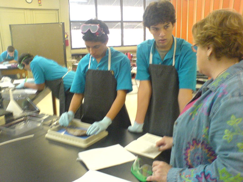
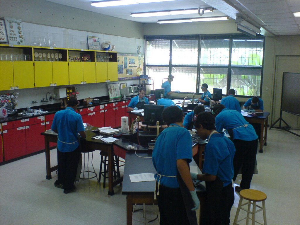
1. Prepare yourself with all the safety materials.
2. Put the crayfish in your dissection pan and pour water to clean the crayfish.
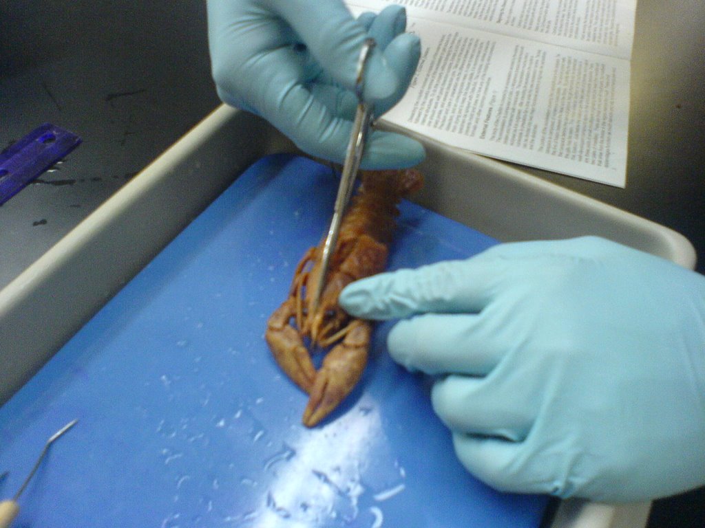
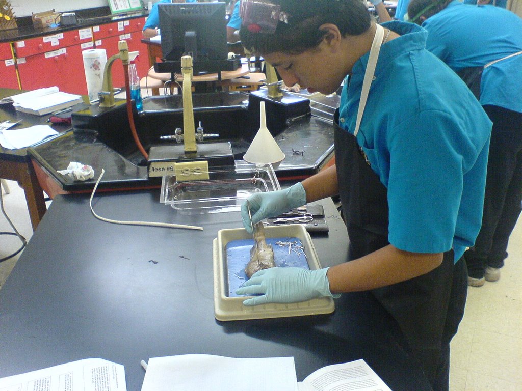
3. Start removing the carapace as follows:
• Carefully insert your scissors under the lower, caudal edge of the carapace and cut dorsally until you have reached the top of the underlying gills lifting the carapace gently with forceps as you go.
• Make an anterior cut in the carapace about 1.5cm to one side of the medial line. Cut forward to within about 1cm of the eye. Repeat this procedure on the other side.
• Carefully remove the strip of exoskeleton that you have formed moving from the tail toward the head. Extend your cuts up so that they join between the eyes.
• Finish removing the dorsal section of the carapace.
• Repeat this procedure one segment at a time on the abdominal segments.
4. Lay your specimen on one side of the dissection tray. Remove the gills.
5. With scissors, cut away the body wall just above the appendage joints.
6. Cut caudally from the first limb joint to the first abdominal segment.
7. From this point, cut up dorsally to the dorsal cut.
8. Cut the cranial end just behind the eye to the mouth.
9. Remove the exoskeleton pieces you have freed.
10. Identify the internal anatomy of the crayfish.
11. When you finish this, dispose of the animal and clean and put in place all the materials.
Observations:
1. We observed that the exoskeleton was a hard material, even though the organism is submerged in water all of its life.
2. One day later this exoskeleton became harder because it was dryer and it was a pale yellow color instead of a vivid orange one.
3. Most organs are the same color, no different ones were noticed between the organs the organism contained, they were all a tan color. The same can be said for the color of the muscles, a brown color identified all of them.
Internal and External Anatomy:
External:
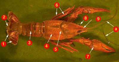
Dorsal View:
1. Uropods, two pairs
2. Telson, the posterior extension of the last body segment. It, in combination with the uropods, is used in rapid, backwards escape swimming.
3. Abdomen, its paired appendages are the swimmerets and the uropods.
4. Cephalothorax, it is covered with a heavily mineralized carapace that offers some protection form predators.
5. Cephalic groove, a lateral seam in the carapace between the head and thorax regions.
6. Walking legs. Each walking leg has an attached gill that occupies the animal's branchial chamber.
7. Cheliped. These appendages are used for defense and food handling.
8. Eye
9. Rostrum, part of the carapace.
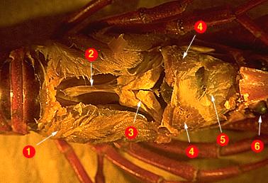
Internal:
Dorsal Cephalotorax View:
1. Gills
2. Heart
3. Hepatopancreas Gland
4. Mandibular Abductor Muscle
5. Gastric Stomach
6. Eye
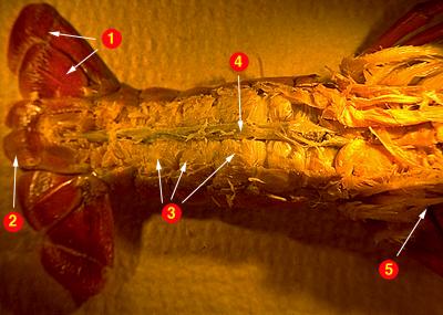
Internal:
Dorsal Abdomen View:
1. Uropods
2. Telson
3. Abdominal Flexor Muscles
4. Intestine
5. Brachial Chamber
Taxonomy:
KINGDOM: ANIMALIA
PHYLUM: ARTHROPODA
CLASS: CRUSTACEA
ORDER: DECAPODA
FAMILY: CAMBARIDAE
GENUS: ORCONECTES
SPECIES: SHOUPI
SCIENTIFIC NAME: ORCONECTES SHOUPI
Conclusion:
The purpose of this laboratory was accomplished, the different internal and external parts of the Crayfish were identified and in the process the use of dissecting materials was enhanced. It was very interesting to see how different animals compared to those of the other groups, not all organisms have the same anatomy or symmetry. It is recommended that for better analysis of the organism, more than one patient dissection is performed and some organs removed in each one to reveal all of the anatomical parts, internal and external.
Bibliographies:
• Hobbs, Michael (1996, March 13). Species NASHVILLE CRAYFISH.
Retrieved November 29, 2006, Web site: http://fwie.fw.vt.edu/
WWW/esis/lists /e454002.htm
• Glase, John C. (2000). Crayfish Review. Retrieved November 29,
2006, Web site: http://biog-101-104.bio.cornell.edu/
bioG101_104/tutorials/animals/crayfish.html